Bone erosion seen in over half of women with Sjögren’s: Study
Volume of bone tissue loss not associated with disease activity
Written by |

More than half of women with Sjögren’s disease show loss of bone tissue, or bone erosion, according to a study carried out in Brazil.
Findings also showed the volume of bone erosion increased by 7% per year, and was 3.5 times higher for women who had gone through menopause. However, the presence of bone erosions or the volume of lost bone was not associated with disease activity, damage scores, or functional disability.
“The findings of this study demonstrate a high prevalence of joint erosions in patients with [Sjögren’s disease], challenging the traditional view of [Sjögren’s] as a non-erosive disease,” the researchers wrote.
The study, “Associations of local bone involvement with disease activity, damage and functional disability in Sjögren’s disease: A cross-sectional study,” was published in Seminars in Arthritis and Rheumatism.
Sjögren’s disease is caused when the immune system mistakenly launches an inflammatory attack against the glands that produce tears and saliva, although other tissues and organs may also be affected. This leads to the hallmark Sjögren’s symptoms, which include of eye and mouth dryness and joint inflammation that may cause bone erosion.
Observational study is 1st of its kind
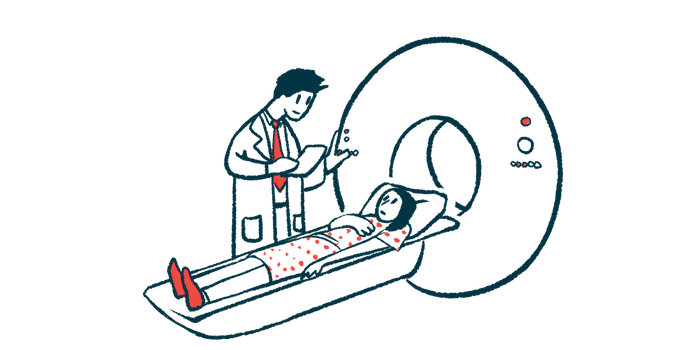
Researchers in Brazil conducted an observational study to assess the relationship between bone erosions and clinical parameters of Sjögren’s using high-resolution peripheral quantitative computed tomography (HR-pQCT). “This is the first study to assess joint and bone involvement in a large cohort of patients with [Sjögren’s disease] using a highly sensitive bone assessment technology,” they wrote.
“Although joint erosions are common, they do not appear to be clinically relevant,” the researchers wrote. “Nevertheless, patients with [Sjögren’s disease] do develop these erosions, and there is likely to be an inflammatory joint component that may be associated with other factors such as age, comorbidities, or poorer disease control.”
A total of 106 women with a mean age of 49.6 who had been living with Sjögren’s for a median of seven years were involved in the analysis.
Of the 100 patients who underwent high-resolution musculoskeletal ultrasound in the hands and wrists, 58% showed signs of inflammation in the joint membrane, while 20% had inflammation in the tissues that protect tendons.
More than half (56.7%) of the patients assessed for joint involvement had bone erosions in the joints of the fingers, with a mean volume of lost bone of 5.71 mm3. For reference, in a group of 60 healthy women, who had been matched for age, sex, and body mass index (BMI; a measure of body fat based on height and weight), the mean volume of lost bone was of 0.38 mm3.
Some 7.2% of the patients had osteophytes, or bone spurs, in finger joints.
Overall, patients with bone erosions were significantly older and had higher levels of vitamin D than those without bone loss.
There were no differences between the two groups regarding BMI, disease duration and activity, menopause, other clinical parameters, medications used, and systemic bone involvement, including bone mineral density, structure, and strength.
Among patients with erosions, the volume of lost bone was correlated with age, BMI, time since diagnosis, disease disability and damage scores, blood sugar levels, and the levels of a marker of bone loss called CTX.
The median volume of lost bone was higher in women who had gone through menopause (8.8 mm3 vs. 1.5 mm3), in those with higher damage (6.1mm3 vs. 2.1 mm3), and in those with osteophytes (28mm3 vs. 2.9 mm3).
“This study shows a higher prevalence of bone erosions in [Sjögren’s disease] than previously described, particularly in older, postmenopausal women,” the researchers wrote, adding that the findings “highlight the potential of HR-pQCT as a sensitive tool to detect joint damage in [Sjögren’s disease].”


